Basic HTML Version


25
Mineralization of human carotids,
Agnieszka Bieniek et al.
www.angiologia.pl
which develops along with aging and is called calcifica-
tion [3].
The basic pathomorfological changes in sclerosis
are sclerotic lamina (plaques) on the internal artery
membrane. [4]. Atheromatous plaques, on which base
mineral lodgement develops, cause stenosis or can lead to
closure of an artery by thrombus. Mineralization develops
mainly within imperforate segments of arteries. In seg-
ments, where the velocity and the volume of the blood
changes, atheromatous changes cumulatemost commonly.
Examples of this are sites of artery ramification. In the case
of carotids, mainly places of ramification of common carotid
artery to internal and external carotid.
Damage to vessel walls and the formation of ath-
eromatous plaques bring about perfect conditions for
mineralization development. Moreover, inflammatory
states, electrolytic disorders, and genetic factors are
favourable to this process [1, 5, 6].
Stenosis and occlusion of the carotid artery are
crucial reasons for central nervous system ischaemia.
Sclerosis is, in 90% of cases, the reason for stenosis and
occlusion of the internal carotid artery. Atheromatous
plaques are usually placed at ramifications of common
carotid artery and at initial segments of external and
internal carotid arteries [6–9].
Reduction of the blood flow through any of carotid
arteries can cause partial cerebral ischaemia by this
carotid. The same result is observed after avulsion of
thrombus from atheromatous plaques and their trans-
mission with blood flow to the brain [6].
The aim of this research was to indicate the mineral
composition of carotid sclerosis lodgement in a group of
patients in which critical narrowing of carotids reducing
blood vessel lumen by 75–80% was diagnosed.
Material and methods
The material for the research was gathered from
a group of 43–71-year-old patients, 6 women (average
age: 67 years) and 20 men (average age: 61.3 years).
All of them had atheromatosis. In all of the patients
critical narrowing of the carotids was recognized. The
narrowing reduced blood vessel lumen by 75–80%.
The patients were qualified to operation of thrombo-
endarterectomy methodology. Mineral-organic sclerosis
lodgement removed during surgery was kept in formalin
(Figure 1–4). In 13 of 26 samples mineral grains were
observed. From the received material, crystals were
separated (with a scalpel and tweezers) and then dried.
The grains were not bigger than 3 mm. Consequently,
examination with polarization microscope was rejected.
The examination was only conducted with a Scanning
Microscope coupled with an Energy Dispersive Spec-
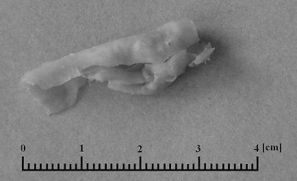
Figure 1.
Sclerosis lodgement removed from carotid.
A 63-year-old woman
Rycina 1.
Złóg miażdżycowy usunięty z tętnicy szyjnej. Kobieta
63 lata
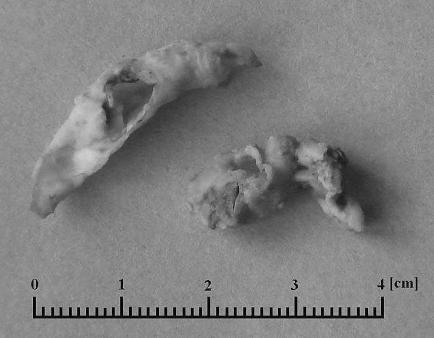
Figure 2.
Sclerosis lodgement removed from carotid.
A 65-year-old woman
Rycina 2.
Złóg miażdżycowy usunięty z tętnicy szyjnej. Kobieta
65 lat
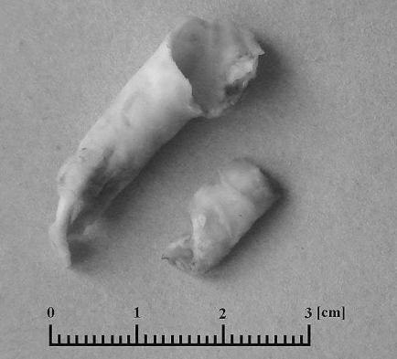
Figure 3.
Sclerosis lodgement removed from carotid.
A 62-year-old man
Rycina 3.
Złóg miażdżycowy usunięty z tętnicy szyjnej.
Mężczyzna 62 lata