Basic HTML Version


20
Acta Angiol, 2012, Vol. 18, No. 1
www.angiologia.pl
of the stent-graft had to begin just below the orifice of
the common carotid artery. Directly after stent-grafting
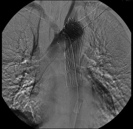
digital subtraction angiography (DSA) was performed
to examine the location and tightness of the prosthesis
(Figure 1), and after 12 days an angio-CT scan was per-
formed to check the position of the graft and whether
there had been an endoleak (Figure 2). The following
radiation dose parameters were calculated: total air
kerma was 3108.4 mGy and DAP was 1137.7 Gy cm².
The cumulative fluoroscopy time was 41 min and the
exposure 2389 ms (in 19 runs, 89 kV, 3 FPS). Total
dose including CT scans exceeded 3Gy. In the first
postoperative day the fall of lymphocyte level was
observed from 30% to 5 %. Peripheral blood swap
parameters normalized after 5 days achieving a lym-
phocyte level of 31%. On the second day a post-op
skin rash was fond on the chest and back. The patient
was placed under dermatological observation and the
rash vanished without treatment after 20 days. Allergic
reaction was excluded after dermatological examina-
tion and the rash was attributed to the radiation dose.
The patient is followed-up in a regular manner every
6 months with special regard to both stochastic and
deterministic effects of radiation. Control CT scans
are followed by peripheral blood examination and
dermatological assessment.
Discussion
Air kerma (Gy) and dose-area product (DAP) (Gy
cm²) are the main parameters measured directly during
various radiographic procedures [6]. In the presented case
of descending aorta stent-graft implantation, the patient
received a very high total dose of radiation because of the
long-lasting procedure. Total air kerma was 3108.4 mGy
and DAP was 1137.7 Gy cm² while the mean DAP value
for this kind of treatment is about 400 Gy cm² [7]. The
cumulative fluoroscopy timewas 41min and the exposure
2389 ms (in 19 runs, 89 kV, 3 FPS).
In this case thoracic aortic endograft implantation
was prolonged because of difficulties with passing
a guidewire into the true lumen so that a stent-graft
could be deployed correctly.
It was not possible to reach
the true lumen via femoral access, so the brachial access
was used. Also, the close location of the dissection flap
in relation to the left common carotid artery made it
necessary to use a guidewire placed inside the carotid
artery via brachial access as a safety measure. Owing to
the unfavourable location of the aneurysm, including the
left subclavian artery, the endograft had to be positioned
very precisely before its deployment, in order not to
cover the left common carotid artery. However, in this
and many other cases the covering of the subclavian ar-
pigtail z prawej tętnicy ramiennej do właściwego światła
aorty. Aby uzyskać przepływ krwi w obu tętnicach bio-
drowych, wykonano fenestrację wewnątrznaczyniową
w odcinku poniżej tętnic nerkowych. Po uzyskaniu do-
stępu do światła właściwego od strony tętnicy udowej po
fenestracji wprowadzono stentgraft do aorty piersiowej
na prowadniku Amplatz 0,035 (Cook Inc USA; Teru-
mo Corp, Tokio, Japonia). Ze względu na objęcie roz-
warstwieniem odejścia lewej tętnicy podobojczykowej
zdecydowano o jej przykryciu protezą. Zakotwiczenie
proksymalnej części stentgraft znajdowało się tuż poniżej
odejścia lewej tętnicy szyjnej wspólnej. Bezpośrednio po
implantacji protezy wewnątrznaczyniowej wykonano
cyfrową angiografię subtrakcyjną (DSA) w celu zbadania
położenia i szczelności protezy (ryc. 1) a po 12 dniach
TK z programem angio w celu wykluczenia migracji
stentgraftu i obecności przecieku (ryc. 2). W trakcie
operacji rutynowo określono dawkę promieniowania:
całkowita Air Kerma — 3108,4 mGy i DAP (
dose area
product
) — 1137,7 Gy cm². Całkowity czas fluoroskopii
wynosił 41 min, a ekspozycja — 2389 ms (w 19. serii,
89 kV, 3 FPS). Całkowita dawka promieniowania, w tym
tomografia przed- i pooperacyjna, przekroczyła 3Gy.
W pierwszym dniu po operacji stwierdzono spadek
poziomu limfocytów z 30% do 5%. Parametry mor-
fologii krwi obwodowej w rozmazie ręcznym uległy
poprawie po 5 dniach, gdy poziom limfocytów osią-
gnął 31%. W 2. dobie pooperacyjnej zaobserwowano
wsypkę na skórze klatki piersiowej i plecach. Pacjent
został poddany kontroli dermatologicznej, a wysypka
ustąpiła bez leczenia po 20 dniach. Reakcję alergiczną
Figure 1.
Intraoperative DSA after stentgraft implantation
Rycina 1.
Śródoperacyjny obraz DSA po wszczepieniu
stentgraftu