IMAGE IN CARDIOVASCULAR MEDICINE
Cardiology Journal
2022, Vol. 29, No. 6, 1043–1044
DOI: 10.5603/CJ.2022.0110
Copyright © 2022 Via Medica
ISSN 1897–5593
eISSN 1898–018X
Added value of contrast echocardiography for the evaluation of multiple giant coronary artery aneurysms with coronary to pulmonary arterial fistulas
Yiwei Zhang12*Ziming Zhang12*Zhenxing Sun12*Yuji Xie12*Yihan Chen12He Li12Lingyun Fang12Li Zhang12Yuman Li12Mingxing Xie12
1Department of Ultrasound, Union Hospital, Tongji Medical College, Huazhong University of Science and Technology, Wuhan, China
2Hubei Province Key Laboratory of Molecular Imaging, Wuhan, China

Address for correspondence: Mingxing Xie, MD, PhD, 1277 Jiefang Avenue, Wuhan, 430022, China, tel: 86-2785726430, fax: 86-2785726386, e-mail: xiemx@hust.edu.cn; or Yuman Li, MD, PhD, 1277 Jiefang Avenue, Wuhan, 430022, China, tel: 86-2785726430, fax: 86-2785726386, e-mail: liym@hust.edu.cn; or Li Zhang, MD, PhD, 1277 Jiefang Avenue, Wuhan, 430022, China, tel: 86-2785726430, fax: 86-2785726386, e-mail: zli429@hust.edu.cn
Received: 4.01.2022 Accepted: 5.09.2022
*These authors contributed equally to this work.
This article is available in open access under Creative Common Attribution-Non-Commercial-No Derivatives 4.0 International (CC BY-NC-ND 4.0) license, allowing to download articles and share them with others as long as they credit the authors and the publisher, but without permission to change them in any way or use them commercially.
A 55-year-old man was admitted to our hospital for transient headache. Twelve-lead surface electrocardiogram (ECG) revealed rapid atrial fibrillation and premature ventricular beat. Transthoracic echocardiography revealed a huge hypoechoic mass adjacent to the aorta and two cystic masses located in the left anterolateral aspect of the pulmonary artery (PA), which were initially suspected as pseudoaneurysms of PA (Fig. 1A, B). Color Doppler flow imaging showed multiple bilateral flows between masses and PA (Fig. 1C). Subsequent contrast echocardiography was performed, which displayed no origination of masses from the aorta or the PA, suggesting giant coronary artery aneurysms (Fig. 1D, E).
In addition, a great quantity of intraluminal thrombi was evident on contrast echocardiography. Furthermore, contrast agent provided better delineation of tortuous coronary arteries with fistulas to the PA (Fig. 1F). Three-dimensional volume-rendered reconstruction images of cardiac coronary computed tomographic angiography (CTA) demonstrated multiple giant coronary artery aneurysms originating from the right coronary artery (RCA) and the left anterior descending artery (LAD) with bilateral coronary artery fistulas to the PA (Fig. 1G, H).
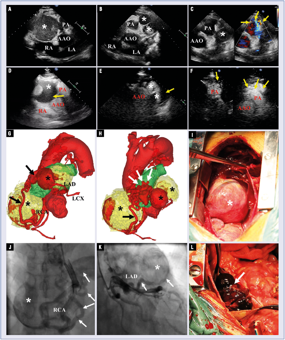
Figure 1. A, B. Two-dimensional echocardiography showing a huge hypoechoic mass (asterisk) adjacent to the aorta and two cystic or solid-cystic masses (asterisk) located in the left anterolateral aspect of the pulmonary artery (PA); C. Color Doppler flow imaging demonstrating a continuous shunt from the coronary artery into the PA (yellow arrows); D, E. Contrast echocardiography clearly displaying the giant coronary artery aneurysm with intramural thrombus (yellow arrows); F. Contrast echocardiography confirms the communication between the tortuous coronary arteries (yellow arrows) and the main PA; G, H. Three-dimensional volume-rendered reconstruction images of cardiac computed tomographic angiography shows multiple giant aneurysms of coronary arteries with intramural thrombi (asterisk), connected by fistulas (black arrows) to the PA (white arrows). (Yellow color indicates aneurysmal thrombus); J, K. Coronary angiography showing the huge coronary artery aneurysms (asterisk) with fistulas from the right coronary artery (RCA) and left anterior descending artery (LAD) to the PA (white arrow); I, L. Intraoperative photographs displaying the huge aneurysm of RCA (asterisk) and thrombus within the aneurysm of LAD (white arrow); AAO — ascending aorta; LA — left atrium; RA — right atrium; LCX — left circumflex.
Invasive coronary angiography confirmed the presence of huge coronary artery aneurysms arising from the RCA and the LAD, associated with coronary-pulmonary fistulas (Fig. 1J, K). During operation, large aneurysms with intramural thrombus arising from the LAD and the RCA and coronary artery fistulas to the PA were observed (Fig. 1I, L). The aneurysms were resected; orifices of fistulae were closed, and the RCA and the LAD were reconstructed. The patient recovered well after the surgery.
This work was supported by the National Natural Science Foundation of China (No. 81727805; 81922033; 81401432).

